


Chordom Cancer, Childhood Treatment In India
Introduction:
Chordomas are rare tumors that arise from embryonic notochordal remnants along the length of the neuraxis at developmentally active sites. These sites are the ends of the neuraxis and the vertebral bodies. Chordomas comprise less than 1% of CNS tumors, though they can occur in extraaxial locations. Chordomas are thought to arise from ectopic notochord remnants.
History of the Procedure
In 1857, Virchow originally described chordomas when he named them ecchondrosis physaliphora, believing they were cartilaginous in origin. In 1895, Ribbert pierced a nucleus pulposus and found similar tumors. From this bit of evidence, he correctly surmised the notochordal origin of chordomas.
Problem
Ecchordosis physaliphora is a term that refers to small, well-circumscribed, gelatinous masses adherent to the brainstem. Although composed of notochordal remnants, ecchordosis physaliphora seldom, if ever, progresses into chordoma. Ecchordosis physaliphora is a reported finding in approximately 2% of autopsy examinations, but chordomas are quite rare. Even though chordomas usually are slow-growing tumors, they are locally aggressive with a tendency to infiltrate into adjacent tissues and organs. Local recurrence results in tissue destruction and generally is the cause of death. Metastases are recognized but are uncommon.
Frequency
Chordomas are rare neoplasms. As primary intracranial neoplasms, they only constitute 0.2% of all CNS tumors; however, they constitute 2-4% of all primary bone neoplasms. Chordomas generally occur in 3 locations, which are, in descending order of frequency, the sacrum, intracranially at the clivus, and along the spinal axis. Fifty percent of chordomas occur in the sacrum, and spinal axis chordomas are rare. Occasional parasellar and sellar examples have been described, and extraaxial sites have been reported in the literature.
 When considering all locations, the male-to-female ratio is 2:1. However, skull base tumors, as a subgroup, tend to have a more equal sex distribution. A number of reports indicate that chordomas are seen in all age groups, with the peak incidence varying by site. Intracranial chordomas present in a much younger age group than their spinal counterparts because the relevant anatomy of the clival region produces earlier symptomatology. In one series of chordomas reviewed, the average age at diagnosis of all patients with chordomas was 56 years, with an age range of 27-80 years. When considered by site, the average age for intracranial chordomas is 48 years; as a subgroup, chordomas of the sphenoccipital area have an average occurrence age of 38 years. The average age for sacrococcygeal chordomas is 56 years. For chordomas occurring along the vertebrae, the average age is 46 years.
Etiology
Chordomas are thought to arise from primitive notochordal remnants along the axial skeleton. During development, the notochord is surrounded by the developing vertebral column. In adults, remnants of the notochord are present as the nucleus pulposus of the intervertebral discs. Notochordal remnants that are extradural are most common at the sacrococcygeal region but can be found at any site along the length of the axial skeleton. The distribution of tumors matches the distribution of notochordal remnants.
A genetic basis has been described for some chordomas. However, most exhibit complex abnormal karyotypes including whole or partial losses of chromosomes 3, 4, 10, and 13, gains in chromosome 7, and rearrangements of chromosome 1p.1 All have been implicated in the pathogenesis of chordomas. Also, microsatellite instability resulting from DNA mismatch repair deficiencies has been demonstrated, however, no chordoma-specific translocations have been identified.
Pathophysiology
Chordomas are characterized by slow growth, with local destruction of the bone and extension into the adjacent soft tissue. Very rarely, distant metastases are encountered. These tumors usually have a relatively indolent but prolonged course with multiple local recurrences, and, eventually, they may be responsible for mortality.
Presentation
The clinical presentation is entirely dependent on the location of the chordoma. At the sacrum, common presenting symptoms are back and/or lower extremity pain. About one half of patients with chordomas have autonomic symptoms, particularly rectal dysfunction or urinary incontinence. About one half of patients with chordomas have a palpable sacral mass.
With intracranial tumors, the most common presenting symptoms are diplopia and headache. Neurologic signs also occur in over one half of the patients, primarily as cranial nerve palsies. Palsies of cranial nerve VI and the sensory branch of V are the most common.
Patients with tumors located along lower vertebrae may present with pain, bladder dysfunction, or lower extremity weakness. Patients with tumors located along cervical vertebrae present with hoarseness, dysphagia, and, occasionally, pharyngeal bleeding. Other rare or unique symptoms have been reported but are the exception. The time span from the onset of symptoms to diagnosis averages 10 months.
Indications
Surgical therapy for these tumors is indicated as they continuously grow, albeit slowly, and erode bone and adjacent soft tissue, causing marked destruction of surrounding tissues.
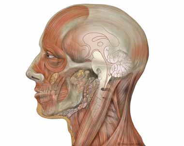
Relevant Anatomy
The location of the notochord along the spinal canal is directly related to the location of notochord remnants, particularly at the ends of the spinal axis. Of chordomas, 49% occur at the sacrococcygeal region, and 30% occur at the sphenoccipital region, with nearly all of these occurring at the clivus. These tumors have a variable extension. Vertebral chordomas account for only 15% of total chordomas and occur in the lumbar, cervical, and thoracic regions in descending order of frequency.
Grossly, chordomas are variable in size. They are soft, gelatinous, smooth, or lobulated and are gray-white in color on their outer surface. On cut section, the tumor is homogeneous in color and consistency. Occasionally, calcifications or hemorrhages are present. Chordomas appear to be encapsulated when in soft tissue but not when they are located in bone.
Contraindications
Contraindications to surgery for excision of a chordoma primarily are related to general health of the patient and preexisting medical conditions. The patient should be evaluated for cardiac, pulmonary, hematological, or endocrine disorders as well as coagulation status. These disorders need to be addressed and managed prior to surgery.
Free Consultation
Conditions


